



Atlas of 6.3 Million Brain Cells Maps Gene Shifts Across Lifespan and Disorders
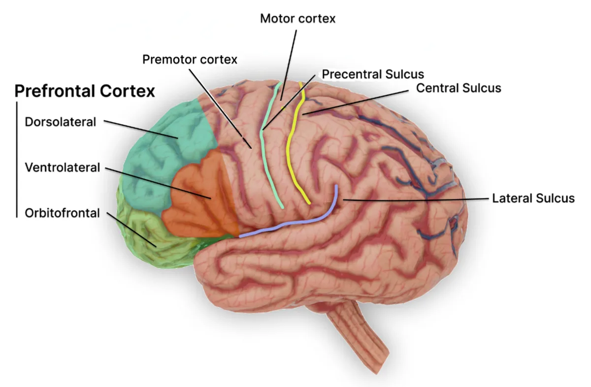
Researchers isolated single nuclei from donated tissue of the dorsolateral prefrontal cortex and ran them through multiplexed single-nucleus RNA sequencing. The method captures the transcriptome of one nucleus at a time: the nucleus is separated, its RNA is converted into a barcoded library, and sequencing then tallies which genes are active inside that cell. Panos Roussos, director of the Center for Disease Neurogenomics at the Icahn School of Medicine at Mount Sinai, steered the PsychAD Consortium effort that produced the maps.
The same region supports working memory, planning, decision-making and cognitive control—functions that falter across many of the conditions the team set out to study. By locking every sample to this one window of cortex and reading gene activity at nuclear resolution, the group could later sort cells by type and compare how their programs shifted with age or diagnosis. Outside the consortium, computational biologist Zhichao Miao of the Guangzhou National Laboratory watched the package land. It arrived as eight or nine companion studies, three of them in Nature.
“The scale of this project and the amount of work required to assemble it are genuinely impressive,” Miao said.
The atlas holds more than 6.3 million individual brain cell nuclei. They came from 1,494 unique donors. Ages stretched from infancy, before birth, to 108 years, and the donors represented diverse genetic ancestries. From the gene-activity patterns inside each nucleus, the team sorted the cells into 8 classes, 27 subclasses and 65 subtypes, among them excitatory and inhibitory neurons, astrocytes, immune cells, oligodendrocytes, oligodendrocyte progenitor cells and vascular support cells.
Cell type accounted for 50.5 percent of the variation in the dataset. Person-to-person differences were the second-largest source. Nuclei from the same donor are related observations rather than independent samples. Jennifer Below, a professor of genetics at Vanderbilt University Medical Center who was not involved in the work but reviewed a companion paper, called the atlas the largest resource with cellular resolution released to date. Comparable single-cell and single-nucleus transcriptomic collections had focused on specific age groups, single disorders or smaller cohorts.
“Single-cell studies of the human brain have traditionally been limited to relatively small numbers of individuals,” Miao said. “Pushing us into a population-scale setting changes the kinds of questions we can ask.”
The PsychAD Consortium was founded in 2019 as an NIH-funded multi-institution partnership, supported by the National Institute on Aging, to link genetic variation, ageing and disease to specific brain cells. Specimens arrived from banks and cohorts whose records had never been built to match: the Mount Sinai NIH Neurobiobank, the NIMH-IRP Human Brain Collection Core, and five prospective cohort studies at the Rush Alzheimer’s Disease Center. Clinical, pathological and demographic metadata differed by source, so the team applied a harmonization scheme that standardized those variables across every donor list.
The consortium held to a single anatomical target, the dorsolateral prefrontal cortex, chosen for its roles in working memory and executive function. Holding that region fixed across ages and diagnoses made the comparisons consistent and tied the new maps to genetic and molecular studies already anchored in the same tissue.
“Studying the same region under different conditions enables more consistent comparisons and helps connect our results to existing genetic and molecular studies,” Roussos said. “This is an important window into brain disease, but additional regions will be needed to understand the full picture.”
From healthy donors the same maps then traced how gene activity itself moves across a lifetime.
From the healthy-donor foundational work—brains collected from individuals between less than one and 97 years old—gene activity resolved into three phases. Early life brought rapid remodeling, with extensive developmental changes related to neurogenesis and gliogenesis, the formation of new neurons and glial cells. Stability held from around age 24 through midlife. Renewed remodeling began around age 65, or near 60 by some accounts, particularly in glial support and immune cells, and linked to immune activity, stress responses and the brain’s circadian rhythms. Different cell types kept distinct patterns of gene activity across the arc.
Most cellular populations in the dorsolateral prefrontal cortex stabilized at approximately age 24.
“It does not mean the brain suddenly finishes developing on someone’s 24th birthday, or that decline begins at that age. Other aspects of brain biology continue to change throughout life,” Roussos said. “This provides a reference for distinguishing typical aging from disease-associated changes.”

Michael Lutz, a professor of neurology and pathology at Duke University who was not involved in the work but reviewed the paper, noted that gene-expression patterns change nonlinearly across disease progression and can even shape its course.
Donors diagnosed with Alzheimer’s disease, dementia with Lewy bodies—listed as diffuse Lewy body disease—Parkinson’s disease, vascular dementia, tauopathy, frontotemporal dementia, schizophrenia and bipolar disorder supplied the disease half of the atlas. The cohorts counted 500 with Alzheimer’s, 112 with diffuse Lewy body disease, 85 with vascular dementia, 40 with Parkinson’s disease, 47 with tauopathy, 15 with frontotemporal dementia, 120 with schizophrenia and 55 with bipolar disorder. Many carried more than one diagnosis.
The team compared transcriptomic variation across the eight conditions, first isolating a universal cross-disease signature and only then the patterns unique to each. Shared changes touched basic cellular functions. After those common threads were discounted, Alzheimer’s, diffuse Lewy body disease, vascular dementia and Parkinson’s showed the strongest remaining genetic and transcriptomic concordance.
“Together, these studies help explain where disease-related changes occur and which biological processes deserve closer investigation,” Roussos said. “A useful treatment needs to influence the right biological process in the right cells. This map helps narrow that search. It can identify vulnerable cell populations, reveal processes associated with preserved brain function and help researchers decide which potential treatment targets to test.”
For donors with Alzheimer’s disease the researchers constructed individual molecular profiles. The profiles revealed person-to-person variation in gene regulation and in projected cell-type interactions, differences present even among people who carried the same diagnosis. Some of those donors had maintained cognitive health despite substantial Alzheimer’s pathology. Their cells exhibited differences in energy-related cellular processes.
“These provide clues to possible protective mechanisms that need further testing,” Roussos said.
Donor brains collected at different times of death made it possible to reconstruct 24-hour circadian cycles of gene activity—the daily schedule of which genes turn on and off inside prefrontal cells. An analysis drawn from approximately 200 neurotypical adults showed that younger and middle-aged adults kept synchronized daily patterns of clock genes, the genes that organize circadian rhythm, inside their neurons. In older adults, particularly those aged 60 and up, the same clock patterns ran weaker and less synchronized. The stamps of time of death, gathered across the donors, assembled into a single daily cycle of gene activity in the prefrontal cortex.